News
[PUBLICATION] MR Passive Elastography: From Seismic Noise Tomography to Brain Tumors Characterization
Dates
on the November 27, 2025
with Jean-Pierre Remenieras, Guillaume Lacoin, Ilyess Zemmoura, and Laurent Barantin
We are pleased to share our new publication in Journal of Magnetic Resonance Imaging: “MR Passive Elastography: From Seismic Noise Tomography to Brain Tumors Characterization”
Authors: Gwenaël Pagé, Khalil Rachid, Benoît Larrat, Bruno Giammarinaro, Johannes Aichele, Guillaume Lacoin, Ilyess Zemmoura, Franck Mauconduit, Angéline Nemeth, Laurent Barantin, Alexandre Vignaud, Jean-Pierre Remenieras, Stefan Catheline, Jean-Luc Gennisson.
Web link - https://onlinelibrary.wiley.com/doi/10.1002/jmri.70072
This study was led by Jean-Luc Gennisson (#BIOMAPS, CNRS, INSERM, CEA, Université Paris-Saclay) and Stefan Catheline (LabTAU, Inserm U1032) with our colleagues Jean-pierre Remenieras (Université de Tours), Guillaume Lacoin (Université de Tours), Ilyess Zemmoura (Université de Tours, CHRU de Tours), and Laurent Barantin (Université de Tours) from our iBraiN Inserm Lab laboratory — an excellent illustration of transdisciplinary and translational research, bridging engineering, MRI physics, neuroscience, and clinical applications.
What this study shows:
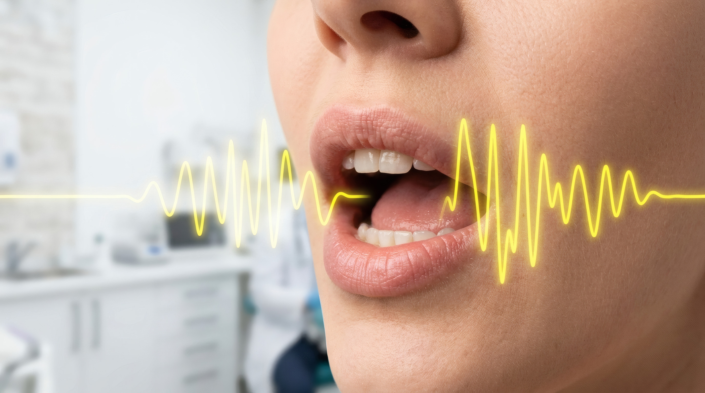
Inspired by seismic noise tomography, the authors adapted the same principles to the human brain, treating it as a system full of natural vibrations (cardiac pulsation, breathing, vocalization, etc.).
Using a modified passive MR elastography (pMRE) sequence at 3T, they evaluated 18 patients with histologically confirmed brain tumors (high-grade gliomas, metastases, low-grade gliomas, meningiomas) and 4 healthy volunteers.
Key findings:
• pMRE-derived shear wavelength strongly correlates with intraoperative ultrasound elastography (r = 0.59).
• By comparing tumor stiffness to surrounding brain tissue, pMRE distinguishes malignant from benign tumors, with excellent performance (AUC = 0.93).
• The technique requires no external mechanical driver, using only intrinsic body vibrations.
Impact
This work demonstrates that intracranial passive MRE is clinically feasible and has the potential to become a powerful tool for:
– early tumor characterization,
– surgical planning,
– treatment monitoring,
– and understanding tumor biomechanics.
A major step toward non-invasive, driver-free MRI-based tumor assessment.
Congratulations to all authors for this groundbreaking contribution.
#MRI #Elastography #Neuroimaging #BrainTumors #TranslationalResearch #SeismicTomography #BiomedicalEngineering #iBraiN #ResearchExcellence
Authors: Gwenaël Pagé, Khalil Rachid, Benoît Larrat, Bruno Giammarinaro, Johannes Aichele, Guillaume Lacoin, Ilyess Zemmoura, Franck Mauconduit, Angéline Nemeth, Laurent Barantin, Alexandre Vignaud, Jean-Pierre Remenieras, Stefan Catheline, Jean-Luc Gennisson.
Web link - https://onlinelibrary.wiley.com/doi/10.1002/jmri.70072
This study was led by Jean-Luc Gennisson (#BIOMAPS, CNRS, INSERM, CEA, Université Paris-Saclay) and Stefan Catheline (LabTAU, Inserm U1032) with our colleagues Jean-pierre Remenieras (Université de Tours), Guillaume Lacoin (Université de Tours), Ilyess Zemmoura (Université de Tours, CHRU de Tours), and Laurent Barantin (Université de Tours) from our iBraiN Inserm Lab laboratory — an excellent illustration of transdisciplinary and translational research, bridging engineering, MRI physics, neuroscience, and clinical applications.
What this study shows:
Inspired by seismic noise tomography, the authors adapted the same principles to the human brain, treating it as a system full of natural vibrations (cardiac pulsation, breathing, vocalization, etc.).
Using a modified passive MR elastography (pMRE) sequence at 3T, they evaluated 18 patients with histologically confirmed brain tumors (high-grade gliomas, metastases, low-grade gliomas, meningiomas) and 4 healthy volunteers.
Key findings:
• pMRE-derived shear wavelength strongly correlates with intraoperative ultrasound elastography (r = 0.59).
• By comparing tumor stiffness to surrounding brain tissue, pMRE distinguishes malignant from benign tumors, with excellent performance (AUC = 0.93).
• The technique requires no external mechanical driver, using only intrinsic body vibrations.
Impact
This work demonstrates that intracranial passive MRE is clinically feasible and has the potential to become a powerful tool for:
– early tumor characterization,
– surgical planning,
– treatment monitoring,
– and understanding tumor biomechanics.
A major step toward non-invasive, driver-free MRI-based tumor assessment.
Congratulations to all authors for this groundbreaking contribution.
#MRI #Elastography #Neuroimaging #BrainTumors #TranslationalResearch #SeismicTomography #BiomedicalEngineering #iBraiN #ResearchExcellence
Contact :
Jean-Pierre Remenieras : jean-pierre.remenieras@univ-tours.fr